How chromosomes shape up for cell division
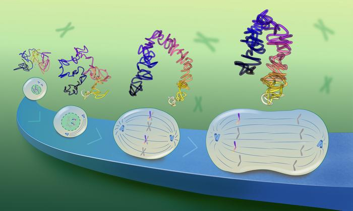
Now, for the first time, EMBL scientists have directly observed this process in high resolution under the microscope using a new chromatin tracing method. The new study shows that the long DNA molecules of each chromosome form a series of overlapping loops during cell division that repel each other. As a result of this repulsion, the DNA loops then stack up to form rod-shaped chromosomes.
Tracing chromosomal DNA in high resolution
Scientists have long hypothesised the importance of DNA loops in building and maintaining chromosomal structure. First identified in the 1990s, condensins are large protein complexes that bind DNA during cell division and extrude it to create loops of varying sizes. Previous studies from EMBL have shed light on the structural mechanics of this process and their essential role in packing chromosomes into forms that can be easily moved between cells.
In fact, mutations in condensin structure can result in severe chromosome segregation defects and lead to cell death, cancer formation, or rare developmental disorders called ‘condensinopathies'.
“However, observing how this looping process occurs on the cellular scale and contributes to chromosome structure is challenging,” said Andreas Brunner, postdoc in EMBL Heidelberg's Ellenberg Group and a lead author of the new paper. “This is because methods for visualising DNA with high resolution are usually chemically harsh and require high temperatures, which together disrupt the native structure of DNA.”
Kai Beckwith – former postdoc in the Ellenberg Group and currently an associate professor at the Norwegian University of Science and Technology (NTNU) – set out to solve this problem. Beckwith and colleagues used a method to gently remove one strand of DNA in cells at various stages of cell division, keeping the chromosome structure intact. They could then use targeted sets of DNA-binding labels to observe the nanoscale organisation of this uncovered DNA strand. This technique, called LoopTrace, helped the researchers directly observe DNA in dividing cells as it progressively formed loops and folds.
“Andreas and I were now able to visualise the structure of chromosomes as they started to change shape,” said Beckwith. “This was crucial for understanding how the DNA was folded by the condensin complexes.”
Loops within loops
From their data, the scientists realised that during cell division, DNA forms loops in two stages. First, it forms stable large loops, which then subdivide into smaller, short-lived nested loops, increasing the compaction at each stage. Two types of condensin protein complexes enable this process.
To understand how this looping eventually gives rise to rod-shaped chromosomes, the researchers built a computational model based on two simple assumptions. First, as observed, DNA forms overlapping loops – first large and then small – across its length with the help of Condensins. Second, these loops repel each other due to their structure and the chemistry of DNA. When the scientists fed these two assumptions into their model, they found that this was sufficient to give rise to a rod-shaped chromosome structure.
“We realised that these condensin-driven loops are much larger than previously thought, and that it was very important that the large loops overlap to a significant extent”, said Beckwith. “Only these features allowed us to recapitulate the native structure of mitotic chromosomes in our model and understand how they can be segregated during cell division.”
In the future, the researchers plan to study this process in more detail, especially to understand how additional factors, such as molecular regulators, affect this compaction process. In 2024, Jan Ellenberg and his team received funding of €3.1 million as an ERC Advanced Grant, to study the folding principles of chromosomes during and following cell division.
“Our newest paper published in the scientific journal Cell marks a milestone in our understanding of how the cell is able to pack chromosomes for their accurate segregation into daughter cells,” said Jan Ellenberg, Senior Scientist at EMBL Heidelberg. “It will be the basis to understand the molecular mechanism of rescaling the genome for faithful inheritance and thus rationally predict how errors in this process that underlie human disease could be prevented in the future.”
In the meantime, a second study from the Ellenberg Team, led by Andreas Brunner and recently published in the Journal of Cell Biology, shows that the nested loop mechanism is fundamental to the biology of cells, and continues during the cell’s growth phase with another family of DNA loop forming protein complexes, called cohesins.
“We were surprised to find that the same core principle of sequential and hierarchical DNA loop formation is used to either tightly pack chromosomes during division into safely movable entities, or to unpack them afterwards to read out the information they contain,” said Ellenberg. “In the end, small, but key mechanistic differences, such as the non-overlapping nature of cohesin-driven loops compared to the strongly overlapping condensin-driven loops might be sufficient to explain the vast differences that we see in the shape the genome takes in interphase and mitosis under the microscope.”
Cell cycle, mitosis, meiosis, cytokinesis, chromatin, chromosome, centromere, spindle fibers, metaphase, anaphase, telophase, prophase, interphase, cell growth, DNA replication, sister chromatids, mitotic spindle, cell differentiation, genetic material, cell regulation
#CellDivision, #Mitosis, #Meiosis, #Cytokinesis, #Chromosome, #DNAReplication, #Genetics, #SpindleFibers, #Interphase, #Prophase, #Metaphase, #Anaphase, #Telophase, #CellCycle, #SisterChromatids, #MitoticSpindle, #CellGrowth, #GeneticMaterial, #CellBiology, #CellRegulation
International Conference on Genetics and Genomics of Diseases
Visit: genetics-conferences.healthcarek.com
Award Nomination: genetics-conferences.healthcarek.com/award-nomination/?ecategory=Awards&rcategory=Awardee
Award registration: genetics-conferences.healthcarek.com/award-registration/
For Enquiries: contact@healthcarek.com
Get Connected Here
---------------------------------
---------------------------------
in.pinterest.com/Dorita0211
twitter.com/Dorita_02_11_
facebook.com/profile.php?id=61555903296992
instagram.com/p/C4ukfcOsK36
genetics-awards.blogspot.com/
youtube.com/@GeneticsHealthcare
Comments
Post a Comment